Published on Dec. 14, 2023
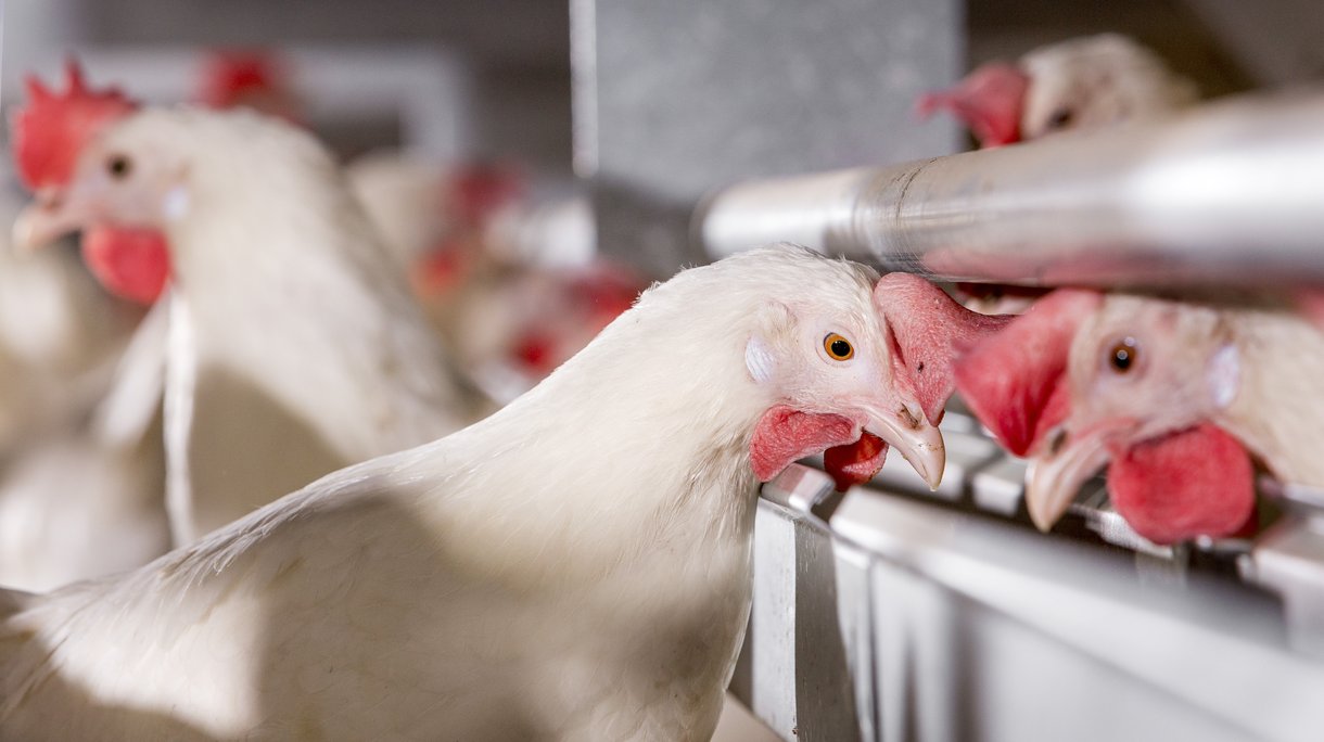
Histomoniasis (Blackhead Disease)
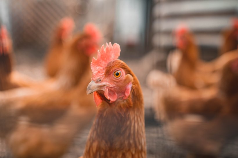
Histomoniasis, also known as blackhead disease, is a parasitic affliction caused by Histomonas meleagridis, an anaerobic protozoan parasite. While chickens typically act as carriers without visible symptoms, the disease proves devastating for turkeys, often resulting in mortality rates between 80% and 100%.
In chickens, the course of the disease is less severe, but it can still cause lesions in the ceca and liver and a significant drop in egg production.. Diagnosis relies on identifying specific cecal ulcerations and necrotic liver lesions. The parasite spreads mainly through cecal nematode eggs, but insects serve as intermediary hosts, facilitating transmission.. Notably, the severity of the infection depends on factors like protozoal genetics, infectious dose, and interactions with cohabiting bacteria and other protozoa in the cecal environment. It’s important to note that, currently, there are no approved treatments or vaccines available for histomoniasis, emphasizing the challenges in managing this disease in poultry populations.
Clinical signs
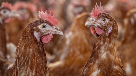
Histomoniasis manifests clinical signs a few days after infection. These signs include listlessness, reduced appetite, drooping wings, unkempt feathers, and the presence of yellow fecal droppings, particularly in the later stages of the disease. Young flocks succumb to the disease rapidly, dying within days after the appearance of symptoms, while older flocks may endure illness for an extended period, becoming weak and decreasing performance before eventual death. Contrary to the name, affected birds do not exhibit a black head. The disease’s name stems from the dark red skin observed in infected birds. As the disease progresses, affected birds exhibit yellow droppings and skin and muscle reddening. The disease develops slowly, leading to significant muscle loss as infected birds become too ill to eat.
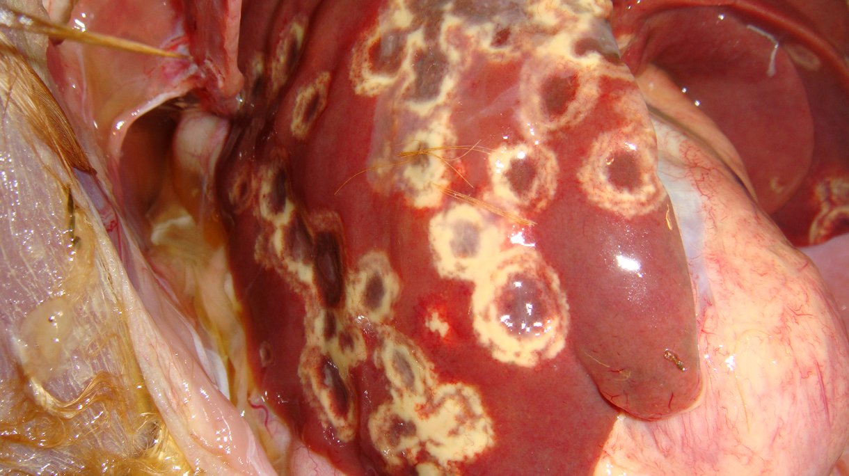
inflammation and the development of a yellowish-green caseous exudate or, in later stages, a dry, cheesy core. These ulcers can sometimes erode the cecal wall, leading to peritonitis and the involvement of other organs. Clinical signs in the ceca become apparent 3-4 days after Histomonas meleagridis invasion. Additionally, histomonas can reach the liver through the vascular system or the peritoneal cavity, leading to the classic and pathognomonic histomonas lesion, a distinct pattern of necrosis of the liver often appearing 6-8 days after infection. These focal liver lesions may be up to 4 cm in diameter and can involve the entire organ, sometimes appearing green or tan. Furthermore, protozoal DNA can be found in other organs, including the kidneys, bursa of Fabricius, spleen, and pancreas.
Diagnosis
Histomoniasis can be diagnosed through several methods. The presence of liver lesions is pathognomonic for this disease. Diagnosis can be confirmed via post-mortem examination, where characteristic lesions are identified in the liver and caecae. Leasions in the caecae need to be differentiated from thyphilitis with coccidiosis. Additionally, histopathologic examination involves analyzing portions of the affected blind gut or liver under a microscope to detect the parasites. PCR (Polymerase Chain Reaction) can be utilized for molecular diagnosis, providing a sensitive and specific method for detecting the disease. However, finding the protoxoa through PCR without the presence of clinical signs is also possible in layer flocks. To distinguish histomonas from other cecal flagellates, scrapings from liver lesions or ceca can be examined microscopically; histomonas are intercellular, although they may appear intracellular due to their close packing. During diagnosis, it’s crucial to differentiate the liver lesions of histomoniasis from those caused by diseases such as tuberculosis, leukosis, avian trichomoniasis, and mycosis. Various diagnostic techniques, including direct examination, histopathology, and molecular methods, are employed to accurately identify and differentiate histomoniasis in affected birds.
Treatment and control
Histomoniasis, presents a significant challenge in poultry, particularly in turkeys, and present even in broiler breeders and layer chickens. Unfortunately, there are no approved drugs for treating this disease. Historically, nitroimidazoles like ronidazole, ipronidazole, and dimetridazole were effective treatments, but they are no longer available. The prevention and control of histomoniasis primarily rely on comprehensive biosecurity measures.. Managing the worm population, ensuring proper manure removal, and deworming birds are vital preventive steps. Oregano essential oil compounds might offer some defense against the parasite and help reduce the risk of blackhead infection. Hygiene is essential; the oral route of infection is common, and the parasite can enter via the vent, necessitating clean living conditions and avoiding ground contamination. Furthermore, avoiding access to feed spilled on the ground and providing clean, dry areas for the birds are vital components of disease prevention. Vaccination against Histomoniasis has been researched but is has not resulted in applicable vaccines.
Despite the absence of specific treatments, effective biosecurity, hygiene practices, and targeted deworming strategies play pivotal roles in minimizing the impact of histomoniasis in poultry flocks.
References
- Beer LC, Petrone-Garcia VM, Graham BD, Hargis BM, Tellez-Isaias G, Vuong CN. Histomonosis in Poultry: A Comprehensive Review. Front Vet Sci. 2022 May 6;9:880738. doi: 10.3389/fvets.2022.880738. PMID: 35601402; PMCID: PMC9120919.
- Daş G, Wachter L, Stehr M, Bilic I, Grafl B, Wernsdorf P, Metges CC, Hess M, Liebhart D. Excretion of Histomonas meleagridis following experimental co-infection of distinct chicken lines with Heterakis gallinarum and Ascaridia galli. Parasit Vectors. 2021 Jun 13;14(1):323. doi: 10.1186/s13071-021-04823-1. PMID: 34120639; PMCID: PMC8201732.
- Dr. Jacquie Jacob. (n.d.). https://poultrydvm.com/condition/blackhead-disease. Retrieved October 5, 2023, from https://poultry.extension.org/articles/poultry-health/common-poultry-diseases/blackhead-in-poultry/
- NADIS. (n.d.). Histomoniasis (Blackhead disease) in Game Birds. NADIS Animal Health Skills. Retrieved October 5, 2023, from https://www.nadis.org.uk/disease-a-z/game-birds/histomoniasis-blackhead-disease-in-game-birds/#:~:text=Blackhead%20disease%20is%20diagnosed%20via,to%20look%20for%20the%20parasites
- Poultry DVM. (n.d.). Blackhead Disease . Poultry DVM. Retrieved October 5, 2023, from https://poultrydvm.com/condition/blackhead-disease
- Robert Beckstead. (2019). Histomoniasis in Poultry. MSD Manual Veterinary Manual. https://www.msdvetmanual.com/poultry/histomoniasis/histomoniasis-in-poultry
- Terra MT, Macklin KS, Burleson M, Jeon A, Beckmann JF, Hauck R. Mapping the poultry insectome in and around broiler breeder pullet farms identifies new potential Dipteran vectors of Histomonas meleagridis. Parasit Vectors. 2023 Jul 20;16(1):244. doi: 10.1186/s13071-023-05833-x. PMID: 37475041; PMCID: PMC10360274.